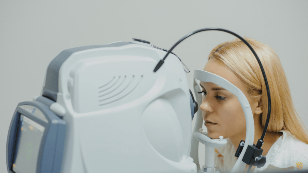
Other than general ophthalmological evaluation, there are certain basic investigations to be done for glaucoma.
Tonometry
Applanation tonometry – gold standard way of measuring the intraocular pressure (eye pressure)
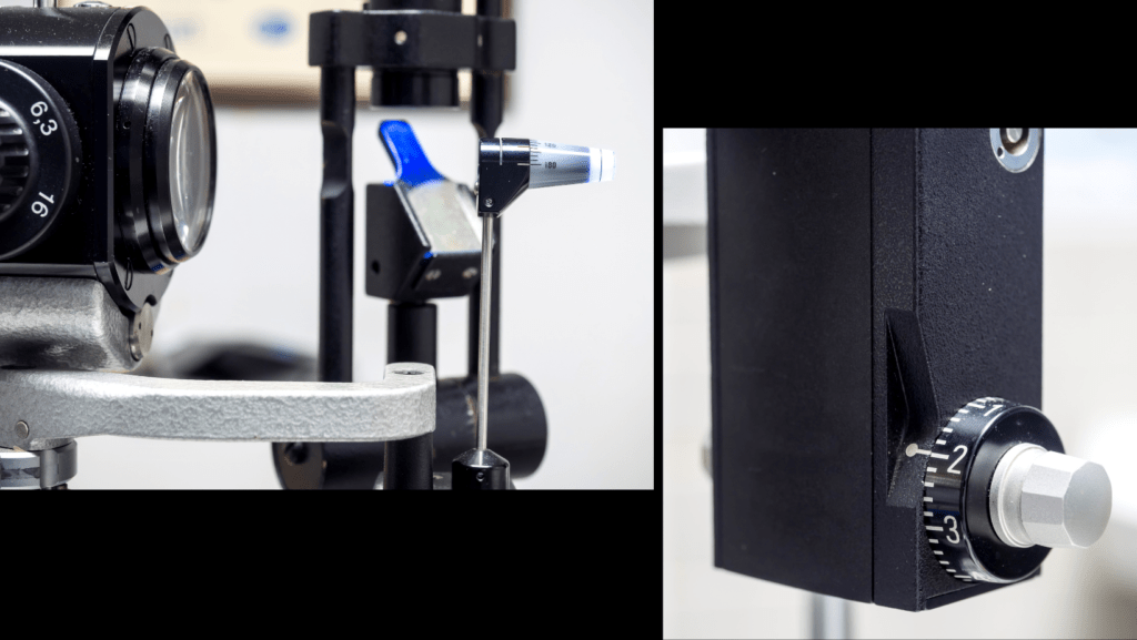

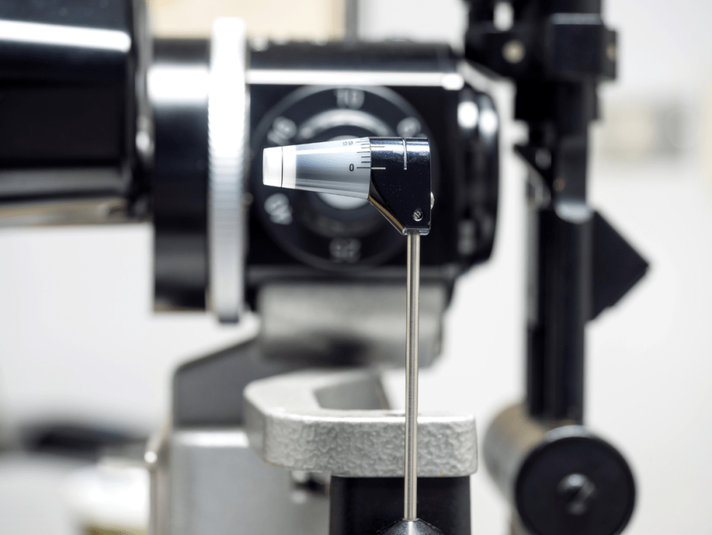

Gonioscope
Using a mirrored lens, the angles of the eye are visualized. It is usually done in a dim room settings.
Follow the link to know – What is the angle of the eye???

Fundus evaluation
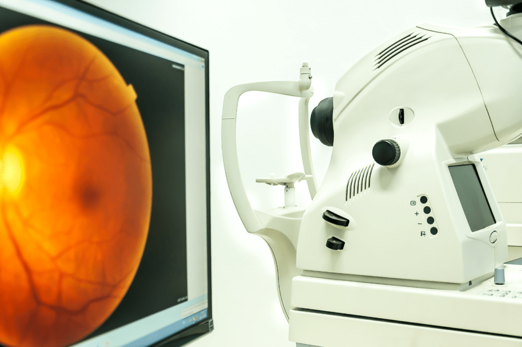
Fundus examination is done to see the retina of the eye. More specifically the optic nerve head which is visualized as a disc is studied during this examination.
Dilatated fundus examination– Eye drops like tropicamide eye drops are placed in the eye and then the fundus is examined after some time. This procedure gives a good 3D view of the optic nerve head for better evaluation.

Fundus Photography
It is the documentation of the fundus examination. A good fundus photograph is an objective representation of glaucomatous changes. Progression of the disease can also be documented with a series of fundus photographs.
Central corneal thickness– Pachymetry
Follow the link to know – What is cornea?
It is the measurement of the thickness of the central part of the cornea. It differs in different individuals.
Significance:
- The actual intraocular pressure measurement depends on the central corneal thickness
- The thin cornea is a risk factor for glaucoma development
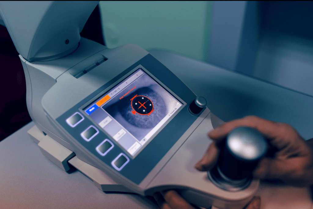
Visual Field test
To know about the visual field – follow the link
This test is done to find out the extent of the visual field loss. It is repeated at certain intervals to compare with the previous reports and identify if there is any progression of the disease.
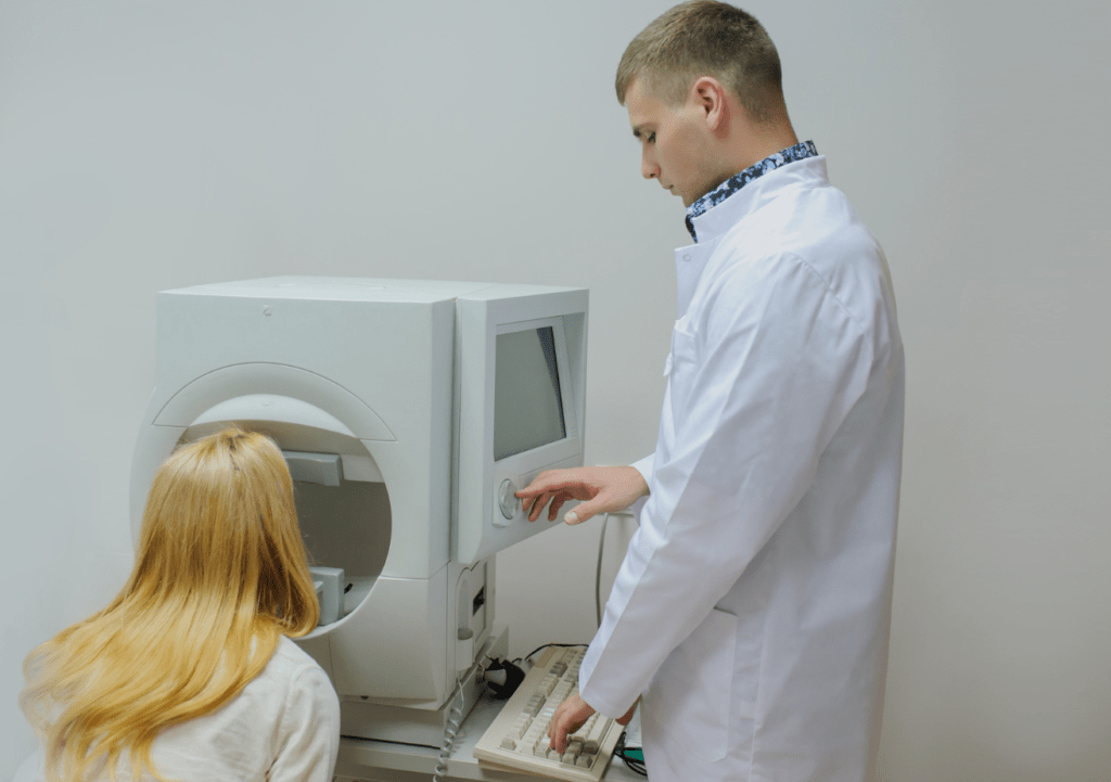
OCT – Optical Coherence Tomography
It is the test done to scan the layers of the retina. It is a non-invasive method. Recent advances in this technology have allowed us to diagnose glaucoma at an early stage. Scan of the optic nerve head and the surrounding area, and the macula have shown to diagnose glaucoma at an early stage. Some softwares can even monitor the progression of the disease.
