The eyeball contains two types of clear fluid. One type of fluid (Vitreous Humour) is stable but the other type of fluid called “Aqueous Humour” (Figure 1) is constantly produced and drained from the eye (Figure 2). This maintains a particular pressure inside the eye called intraocular pressure. This pressure helps maintain the size and shape of the eyeball.
Normal intraocular pressure is different for different individuals. The range is approximately between 10 to 21mmHg. Some individuals can have higher than 21 mmHg or lower than 10mmHg as normal.
Follow the link to know about the basic structure of the eye

So what happens if there is overproduction of the aqueous or more commonly, block in the drainage?
– The fluid inside the eye increases, which increases the pressure inside the eye.
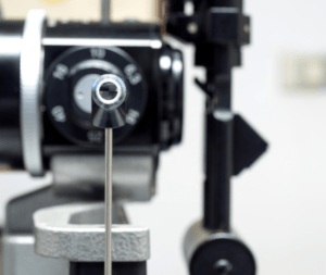
How to measure intraocular pressure?
There are many methods developed to measure the intraocular pressure. The most common ones are
Non contact tonometery- Measuring intraocular pressure (IOP) often involves gently applying a puff of air onto the cornea. While this can briefly surprise or tickle the eye’s surface, most people tolerate it well. Some individuals may feel slight discomfort or a startle reflex due to the suddenness of the air puff, but the procedure is typically quick and not significantly painful

Goldmann applanation tonometry– It involves gently touching the cornea with a small probe. This may cause a slight pressure sensation or a foreign body feeling on the eye. While some individuals may experience mild discomfort or a reflex to blink, the procedure is generally well-tolerated. Anesthetic eye drops are typically used to minimize any potential discomfort. Despite the temporary sensations, Goldmann applanation tonometry is considered a reliable and gold standard technique for accurately assessing IOP.